Anatomy Of Internal Organs Female : Internal Organ Hd Stock Images Shutterstock. Learn now at kenhub their female anatomy diagram: The female reproductive anatomy includes both external and internal structures. The vulva and labia form the. Posted on january 8, 2016 by admin. The vagina is a canal that joins the cervix (the lower part of uterus) to the outside of the body.
The internal genitalia are those organs that are within the true pelvis. The internal female genitalia the internal genitals include paired ovaries and oviducts (fallopian tubes), the uterus, and the vagina. In virgins, the hymen usually encircles the opening like a tight ring, but it may completely cover the opening. The vagina is a canal that joins the cervix (the lower part of uterus) to the outside of the body. Female internal organ with muscles anterior view in this image, you will find larynx, trachea, left subclavian a., the arch of the aorta, pulmonary a., pulmonary trunk, pulmonary v., left atrium, lang, left ventricle, diaphragm, spleen, stomach, transverse colon, jejunum, descending colon, ureter in it.
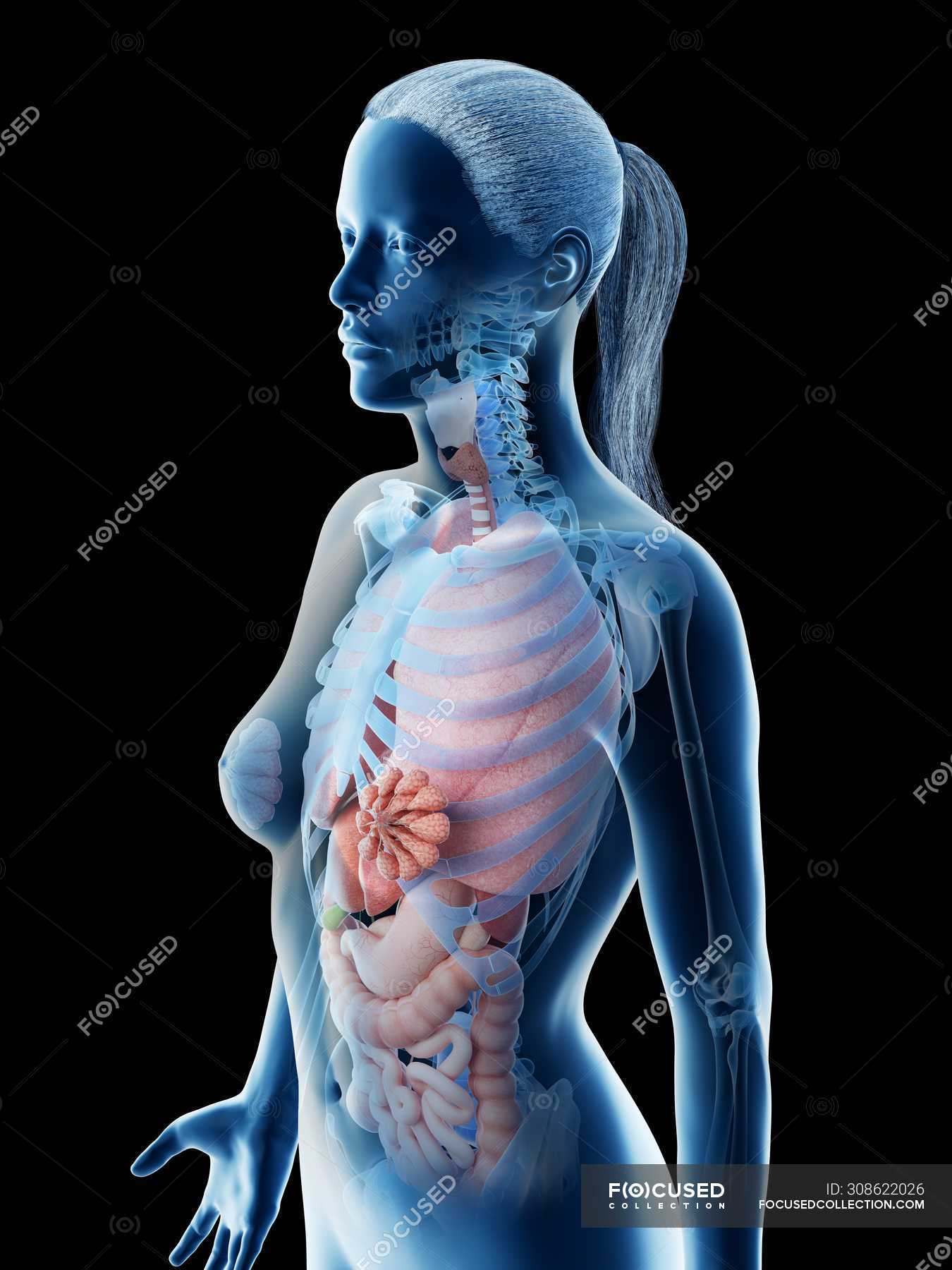
Human Body Model Showing Female Anatomy With Internal Organs Digital 3d Render Illustration Normal Colon Stock Photo 308622026 from st.focusedcollection.com Ovaries, uterine tubes, and uterus. P1y7hg (rf) internal organs of the human icons set, flat style. The female body contains many organs that work together to achieve a variety of functions. This diagram depicts female human anatomy 744×1116 with parts and labels. It extends from the uterus to the vulva (external genitalia). The vulva and labia form the. The external genitalia comprises the structures outside of the true pelvis, including the labia majora and minora, vestibule, bartholin glands, skene glands, clitoris, mons pubis, perineum, urethral meatus, and periurethral area. The vulva is the part of your genitals on the outside of your body — your labia, clitoris, vaginal opening, and the opening to the urethra (the hole you pee out of).
The female reproductive system is an intricate arrangement of structures that can separate into external and internal genitalia.
The female reproductive anatomy includes both external and internal structures. Female genitalia the female sexual organs have reproductive and sexual functions and are divided into internal and external sexual organs. The vagina is an elastic, muscular canal with a soft, flexible lining that provides lubrication and sensation. The vulva is the part of your genitals on the outside of your body — your labia, clitoris, vaginal opening, and the opening to the urethra (the hole you pee out of). One major difference between males and females is their. Browse 21,727 female body organs stock photos and images available, or start a new search to explore more stock photos and images. Kidney transplant in the urology, nice, france, kidney is taken from a living related donor, the recipient's wife transplanting the donated organ the. The vagina meets the external organs at the vulva, which includes the labia, clitoris, and urethra. The female body contains many organs that work together to achieve a variety of functions. The vagina connects the uterus to the outside world. Internal female genital organs the hymen, a mucous membrane, is located at the beginning of the genital tract, just inside the opening of the vagina (see figure external female genital organs). The vulva and labia form the. The vagina is located posterior to the urinary bladder and urethra, and anterior to the rectum.
These organs produce hormones, aid with reproduction or allow for pleasure during sex. • the female reproductive organs can be subdivided into a) external genitalia b) internal genitalia c. Female genitalia the female sexual organs have reproductive and sexual functions and are divided into internal and external sexual organs. One major difference between males and females is their. This diagram depicts female human anatomy 744×1116 with parts and labels.
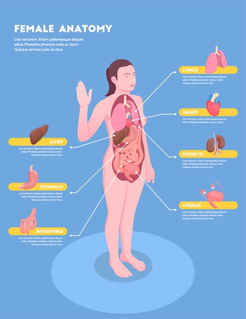
Free Vector Female Anatomy Isometric Infographics With Woman Body And Internal Organs 3d from image.freepik.com The uterus is part of a female's internal anatomy and plays an important role in periods and pregnancy. These organs produce hormones, aid with reproduction or allow for pleasure during sex. Finger pointing to blood vessels. The vagina connects the uterus to the outside world. An illustration showing a theory of vision published in treatise on man by rene descartes. Female internal organ with muscles anterior view in this image, you will find larynx, trachea, left subclavian a., the arch of the aorta, pulmonary a., pulmonary trunk, pulmonary v., left atrium, lang, left ventricle, diaphragm, spleen, stomach, transverse colon, jejunum, descending colon, ureter in it. The internal reproductive organs in the female include: The external genitalia comprises the structures outside of the true pelvis, including the labia majora and minora, vestibule, bartholin glands, skene glands, clitoris, mons pubis, perineum, urethral meatus, and periurethral area.
Ovaries, uterine tubes, and uterus.
The vagina is located posterior to the urinary bladder and urethra, and anterior to the rectum. The internal genitalia are those organs that are within the true pelvis. This diagram depicts female human anatomy 744×1116 with parts and labels. It also is known as the birth canal. The vagina is a canal that joins the cervix (the lower part of uterus) to the outside of the body. Female anatomy of internal organs with skeleton, rear and front views. The external genitalia comprises the structures outside of the true pelvis, including the labia majora and minora, vestibule, bartholin glands, skene glands, clitoris, mons pubis, perineum, urethral meatus, and periurethral area. Functionally, it facilitates menstruation, sexual intercourse and childbirth. It extends from the uterus to the vulva (external genitalia). The uterus is part of a female's internal anatomy and plays an important role in periods and pregnancy. The external female genital (vulva. The vagina connects the uterus to the outside world. The function of the external female reproductive structures (the genital) is twofold:
Anatomy of female reproductive organs by amrit kaur. Acid reflux or heartburn, the photo of stomach and internal organs is on the men's body against gray background, stomach ache, bad health, male anatomy concept. Organs cropped shot of woman holding paper crafted human internal organs on blue background The external female genital (vulva. Kidney transplant in the urology, nice, france, kidney is taken from a living related donor, the recipient's wife transplanting the donated organ the.
Pin On Medical Care Rescue To Hurt Someone Is To Be Strong Yet To Help Them Is To Be Even Stronger from i.pinimg.com Introduction • the reproductive organ in female are those which concerned with copulation, fertilization, growth and development of fetus and its subsequent exit to the outer world. The human female reproductive system (or female genital system) contains two main parts: The internal reproductive organs in the female include: Finger pointing to blood vessels. The vagina meets the external organs at the vulva, which includes the labia, clitoris, and urethra. It has several important functions, including: Collection with heart, liver, lungs, kidneys, stomach, female reproductive system, brain, intestines. Browse 16,500 female human internal organ stock photos and images available or start a new search to explore more stock photos and images.
It also is known as the birth canal.
Organs cropped shot of woman holding paper crafted human internal organs on blue background Collection with heart, liver, lungs, kidneys, stomach, female reproductive system, brain, intestines. The vagina is attached to the uterus through the cervix, while the uterus is attached to. Female internal organ with muscles anterior view in this image, you will find larynx, trachea, left subclavian a., the arch of the aorta, pulmonary a., pulmonary trunk, pulmonary v., left atrium, lang, left ventricle, diaphragm, spleen, stomach, transverse colon, jejunum, descending colon, ureter in it. P1y7hg (rf) internal organs of the human icons set, flat style. The female reproductive organs can be subdivided into the internal and external genitalia (see the images below). The vagina is a canal that joins the cervix (the lower part of uterus) to the outside of the body. The female reproductive system is an intricate arrangement of structures that can separate into external and internal genitalia. To enable sperm to enter the body and to protect the internal genital organs from infectious organisms. Fallopian tubes and ovaries form the adnexa of the uterus. These organs produce hormones, aid with reproduction or allow for pleasure during sex. It has several important functions, including: Learn now at kenhub their female anatomy diagram: